The most powerful flexor of the hip is a single muscle formed from the merging of two individual muscles of the abdomen. Up to 24 cash back 2.
Solved Correctly Label Each Of The 3 Highlighted Muscles Chegg Com
Rib superior to each intercostal muscle.

. Indicate which muscle is highlighted in the image. Anatomy and Physiology questions and answers. Match the infrahyoid muscle to its corresponding image.
Rib superior to each intercostal muscle. 104 Structures of the Muscular System Name __ Handout Date_____ Directions. Correctly label the muscles of the neck back and gluteal region.
The Three Types of Muscle Tissue The body contains three types of muscle tissue. For example a scar in the anterior front carpal wrist. Correctly label the muscles of the neck back and gluteal region.
Each muscle is wrapped in a sheath of dense irregular connective tissue called the epimysium which allows a muscle to contract and move powerfully while maintaining its structural integrity. Classify each of the following terms as a projection P or a depression or opening D. We review their content and use your feedback to keep the quality high.
Identify the highlighted muscle. Identify the highlighted muscle. The cells are striated and multinucleated appearing as long unbranched cylinders.
Select the correct label and characteristic for each of the featured muscles. Up to 24 cash back 1. Describe the action of the highlighted muscle.
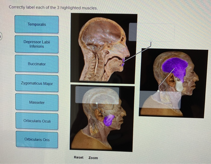
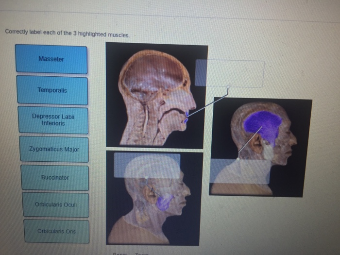
If youre looking for a speedy way to learn muscle anatomy look no further than our anatomy crash courses. Correctly label the muscles of the thoracic cavity and the abdomen. Masseter Temporalis Depressor Labi Inferioris Zygomaticus Major Buccinator ris Oculi.
Muscles of the Body DRAFT. Often these terms are incorporated into the names of muscles that contribute to producing that type of movement at one of the bodys joints. This muscle extends from the back of the knee to.
Outside of upper part of femur. One end is pulled by the muscle to create movement. Extension and lateral rotation of leg.
Each skeletal muscle has three layers of connective tissue that enclose it provide structure to the muscle and compartmentalize the muscle fibers within the muscle Figure 1021. Correctly label each of the 3 highlighted muscles. The three types of muscle cells are skeletal cardiac and smooth.
Group each of the following bones into one of the four major bone cate- gories. Play this game to review Human Anatomy. Identify the highlighted muscle.
It does not matter how the body being described is oriented the terms are used as if it is in anatomical position. Skeletal muscle is voluntary and responds to conscious stimuli. A skeletal muscle b smooth muscle and c cardiac muscle.
Muscles are generally attached at two points in the body. Color the diagrams as you wish. Correctly label each of the 3 highlighted muscles.
Their morphologies match their specific functions in the body. Rib inferior to each intercostal muscle. Abduction and medial rotation of leg.
Correctly label each of the 3 highlighted muscles. Indicate the identity of the two muscles in the images. Rib inferior to each intercostal muscle.
Identify the highlighted muscle. Identify the highlighted muscle. From top LM 1600 LM 1600 LM 1600.
Correctly label each of the 3 highlighted muscles. Inguinal ligament linea alba. The highlighted muscle is the flexor pollicis _____.
Temporalis Depressor Labii Inferioris Buccinator Zygomaticus Major Masseter Orbicularis Oculi Orbicularis Oris Reset Zoom. Identify the highlighted muscle. Mid outer surface of pelvis.
Indicate if the muscle is on the posterior side of the body. Using this standard position reduces confusion. Identify the highlighted muscle.
Cardiac muscle is involuntary and found only in the heart. Sometimes the locations of muscless origins or insertions are incorporated into their names. This is a back view of the right leg.
Preview this quiz on Quizizz. Outside of upper part of femur. Rear part of pelvis sacrum and coccyx.
Identify the type Of muscle in each of the illustrations in Figure 61. The upper limbs are held out to each side and the palms of the hands face forward as illustrated in Figure 141. Identify the highlighted muscle.
Specify each featured eye muscle by selecting its corresponding label. Micrographs provided by the Regents of University of Michigan Medical School 2012. Label the structures of the muscular system.
Anatomy and Physiology questions and answers. Correctly label each of the 3 highlighted muscles. Select the correct label and characteristic for each of the featured muscles Transversus abdominis Insertion.
Muscles of the Body DRAFT. Pubic body linea alba sternum xiphoid process Internal abdominal oblique Origin. Enter the appropriate letter in the answer blanks.
Human body muscle diagrams. One of the large muscles of the leg it connects to the heel. Abduction and medial rotation of leg.
Draw and label the posterior view of a human figure for extra credit. Upper part of pelvis. It flexes and extends the foot ankle and knee.
Movement along superiorinferior axis to bring ribs closer together. Regarding the functions of muscle tissues circle the term in each of the groupings that does not belong with the other terms. What is the name of muscle A.
Gastrocnemius calf muscle. Correctly label each of the 3 highlighted muscles. Studying these is an ideal first step before moving onto the more advanced practices of muscle labeling and quizzes.
Urine Heart bility Foodstuffs Cardiac muscle Smooth muscle. Muscle diagrams are a great way to get an overview of all of the muscles within a body region. Intercalated discs Figure 61 3.
4th - 6th grade. Select the action for which the featured muscle is responsible.
Solved Correctly Label Each Of The 3 Highlighted Muscles Chegg Com

Muscles Worksheet Flashcards Quizlet

Muscle Activation Posture Stretching Pelvic Tilt Muscle Imbalance Posture Stretches
Muscles Worksheet Flashcards Quizlet

0 Comments